

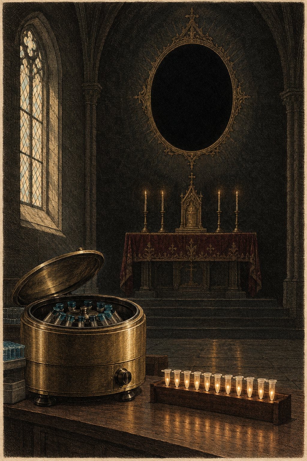
A religion does not need a god. It needs an invisible agent, a priesthood licensed to see it, and a rite that turns doubt into heresy. For a century the agent has been the germ. The priesthood has been the laboratory. The rite, in our time, is a number of cycles.
Nobody in the chain that declared a person a case was asked to see the particle. The public was told the particle was too small for ordinary eyes, that only experts and their instruments could confirm it, and that a positive test was that confirmation. What the test confirmed was not a particle. It was that a short stretch of nucleic acid, copied enough times, crossed a line the laboratory had drawn.
I have been writing toward this for years, from the influenza literature to the exosome question to the measles counts that never received a genotype. This essay is the methodological core those pieces were circling. It is also a correction of a slogan I will not use. The electron microscope is not imaginary. The particle that was supposed to appear in it, again and again, was.
What the Faith Requires
Classical germ theory, in the form that still organizes public health, says a specific invisible particle causes a specific disease, and that finding the particle’s signature is finding the cause. The public cannot check this. The particle is below the reach of a light microscope. Faith is delegated.
That delegation would be science if the delegated observation were the thing claimed: a whole, infectious particle, recovered from the sick, shown to produce the illness, and distinguished from the debris a damaged cell already makes. It is not science when the observation is a manufacturing step, and the manufacturing step is then renamed a diagnosis.
In May 2025 I put the older problem this way: for over a century, germ theory and genetic determinism have cast “invisible enemies — germs and faulty genes — as the primary causes of disease,” and the resulting model has functioned “more as a psychological operation than a scientific certainty” (Genes, Germs, and the Myth of the Invisible Enemy). The XENOGEN frame I offered there was deliberate. It refuses the fake choice between “viruses do not exist” and “a PCR positive is a pathogen.” This essay stays inside that refusal, and looks at the instrument.
The Microscope Was There. The Particle Often Was Not.
Viruses are too small for a light microscope. Transmission electron microscopy is a real method, and it has been used for decades to look at particles in the 50 to 300 nanometer range (a review of RNA-virus replication organelles). Anyone who says the machines do not exist is wrong, and that error hands the argument away.
The live question is different. During the emergency, a clinical case was not an electron micrograph. It was a swab, a cycle count, and sometimes a phone call about symptoms. When researchers later claimed the pictures did show the particle inside human tissue, a multicentre audit in eBioMedicine read 144 publications against structural criteria. Nine presented sufficient structural evidence of virus particles. 135 misinterpreted cellular structures as virus, or showed evidence too poor to decide (Krasemann et al., eBioMedicine, 2022).
What had been called virus was, in the audit’s words, “coated vesicles, vesicles of rough endoplasmic reticulum, multivesicular bodies, and autolytic mitochondria.” Of 337 electron micrographs in those 135 papers, 67 — 20 percent — even had enough structural preservation to judge. The authors’ conclusion is blunt: using refined criteria, 135 of 144 publications “do not sufficiently prove the presence of viral particles in various human tissues.”
Technical note. The same audit did not say no particle has ever been imaged. Nine papers met the bar, and cultured isolates are a different specimen from autopsy tissue. The finding is about the public record of “we saw it in the patient.” Most of that record was the cell, misread.
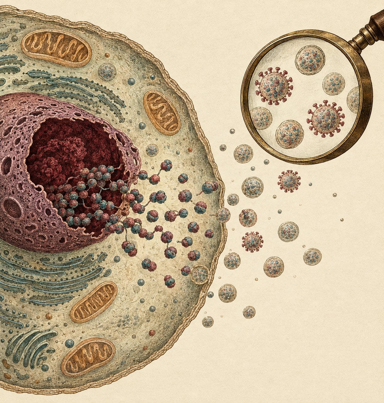
This is the religious structure in laboratory dress. The instrument exists. The claim outruns the instrument. Dissent is treated as denial of the instrument rather than as a demand to see the particle the instrument was supposed to show. A vesicle is not a virus because an expert needed it to be one.
Mullis, Without the Meme
Kary Mullis invented the polymerase chain reaction and received the 1993 Nobel Prize in Chemistry for it. He died in August 2019. He never commented on a COVID test. Anyone who puts a COVID-specific denunciation in his mouth is fabricating a witness.
What he did say, on camera, at a 1997 meeting in Santa Monica about HIV, is enough. Asked how PCR gets misused, he said: “And with PCR, if you do it well, you can find almost anything in anybody.” Later in the same conversation: “[PCR is] just a process that’s used to make a whole lot of something out of something. That’s what it is. It doesn’t tell you that you’re sick, and it doesn’t tell you that the thing you ended up with really was going to hurt you or anything like that” (Full Fact, quoting the 1997 recording).

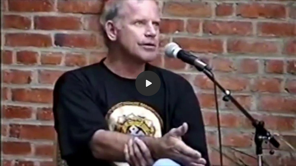
Read that carefully, because the viral meme sanded it down. Mullis did not say the words “anyone can test positive for practically anything if you run it long enough” as a single sentence. Fact-checkers are right about the stitching. They are wrong if they treat the stitching as a reason to ignore the stitches. He said three things that survive the correction:
PCR is a manufacturing process. It makes a lot of a sequence out of a little of a sequence. His Nobel lecture is about that invention, not about diagnosis (Nobel lecture, 1993).
Done well, it can find almost anything in anybody. “Done well” is the point. Sensitivity is the feature. It is also the hazard.
The product does not tell you that you are sick, and it does not tell you that what you amplified was going to hurt you.
That is not a conspiracy theory. It is the inventor describing the resolution of his own tool. PCR identifies neither an infectious particle nor a disease. It reports that a chosen sequence, or something close enough to it, was copyable.
David Crowe put the operational consequence in a piece I published in 2020: the coronavirus test “does not produce a positive/negative result, but simply the number of cycles required to detect genetic material. The division between positive and negative is an arbitrary number of cycles chosen by the testers,” and “positive tests do not mean infected” (GreenMedInfo). I have repeated the distinction since, including on measles: “PCR is considered a sensitive surveillance tool, but sensitivity is not specificity. It detects genetic material, not whether a virus is viable, infectious, or even wild-type” (789 Cases, Zero Genotypes).
The Yield Is the Result
A PCR “positive” is a yield. Each cycle roughly doubles the target. After 30 cycles you are looking at about a billion-fold amplification; after 40, about a trillion-fold. The cycle threshold, or Ct, is the cycle at which the signal crosses the machine’s line. A low Ct means a lot of starting material. A high Ct means the machine had to work a long time to see anything.
The World Health Organization told laboratories this in January 2021, after the emergency had already been governed by the test. “The cycle threshold (Ct) needed to detect virus is inversely proportional to the patient’s viral load.” Most PCR assays, WHO wrote, are “indicated as an aid for diagnosis,” and the result has to be read with specimen type, timing, clinical findings, and exposure history. If the result does not match the patient, take a new specimen (WHO Information Notice for IVD Users 2020/05).
An aid for diagnosis is not a diagnosis. That sentence arrived late, and it did not unwind the year in which a case was the aid.
The culture data show what the aid was aiding.
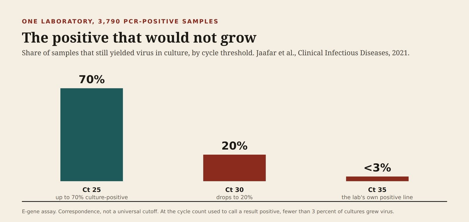
In a correspondence in Clinical Infectious Diseases, Jaafar and colleagues inoculated 3,790 PCR-positive nasopharyngeal samples and recovered 1,941 isolates. At a Ct of 25, up to 70 percent of samples still grew virus in culture. At a Ct of 30, that figure was 20 percent. At a Ct of 35 — the value their laboratory used to call a result positive — fewer than 3 percent of cultures were positive (Jaafar et al., Clinical Infectious Diseases, 2021).
Technical note. This is one laboratory’s E-gene assay, published as correspondence, later corrected on a separate point in the same journal. It is not a universal law of every kit. It is a direct comparison, in thousands of samples, between the thing the public was told (positive) and the thing infectivity requires (virus that grows). The two diverge as the cycle count rises. A religion that calls both results by the same name has stopped doing science.
I wrote in 2024 that the same technology, pointed at avian influenza, “will only serve to raise false case counts” if the COVID lesson is ignored (GreenMedInfo). The lesson is not that every amplification is a fiction. It is that a case defined by yield, without a stated cycle limit and without culture or a full genome, is a case defined by a dial.
What a Damaged Cell Pours into the Blood
Here is the part that gets sloganized, and that I want stated exactly.
When cells die — in sepsis, trauma, ischemia, autoimmune injury, severe inflammation — the nucleus does not stay a locked room. Histones, the proteins that package DNA, are released into the extracellular space in three forms: free, bound to DNA as nucleosomes, or inside neutrophil extracellular traps. All three “can be detected in serum after significant cellular death” (Silk et al., Cell Death & Disease, 2017). Necrosis, the authors write, ruptures the plasma membrane and releases “the intracellular contents of a cell including intra-nuclear proteins.” Once outside, those histones are not inert. They are damage-associated molecular patterns. They activate Toll-like receptors and drive endothelial injury and cytokine release. They can make a person look infected.
Cell-free DNA in the circulation “is released from dying cells” and circulates as nucleosome-sized fragments (Trends in Immunology review). Liquid-biopsy science is built on this fact. The wreckage is real, measurable, and inflammatory by itself.
PCR does not detect those proteins. It detects nucleic acid. That distinction matters, and it does not rescue the test. The same death that dumps histones into plasma dumps the DNA they were wrapped around, mitochondrial nucleic acids, and RNA in extracellular vesicles. The tube is not a clean vial containing one foreign genome. It is a sample from a body already shedding its own nucleus.
This is why I have argued, in the Poisoned, Not Infected series, that the clinical picture called infection can be initiated by toxic and inflammatory injury, and that the narrow choice between “viral contagion” and “terrain” is a false one (the series). Extracellular histones are one mechanism for that picture. A PCR primer run against the nucleic acids those dying cells released is not, by itself, a way out of it.
Homology, and the Resolution PCR Does Not Have
A diagnostic RT-PCR does not read a virus. It copies a short amplicon, often well under 200 base pairs, sometimes nearer 70, chosen because a primer pair sticks to it. That is the resolution of the test. A match at that length is a match at that length. It is not a portrait of a particle, not a proof the sequence was inside a capsid, and not a proof the sequence was infectious.
Three facts limit what such a match can mean.
Shared sequences are normal. Coronaviruses share genome. FDA documents for assays using the CDC N1 primer/probe set report high sequence homology of the N1 probe with SARS-CoV and bat SARS-like coronaviruses, while stating that, combined, the set showed “no significant homologies with human genome, other coronaviruses or human microflora that would predict potential false positive rRT-PCR results” for that design (FDA EUA summary). I am not going to pretend that sentence does not exist. A well-designed primer set can be checked against the human genome and fail to find a predicted cross-reaction. That is a constraint on the claim, not a surrender of it. The check is in silico, primer-specific, and silent about whether a high-cycle signal in a damaged host is a virion.
The human genome is already full of viral-looking sequence. About 8 to 9 percent of it is endogenous retroviral elements (HERV review, 2015). Short-amplicon methods have already been fooled by that fact. The XMRV episode — a claimed human retrovirus that collapsed — was driven in part by primers with nonspecific binding, and later work has shown that only limited stretches of a genome can even distinguish an exogenous retrovirus from an endogenous one. Reconstructing identity from a short amplicon produces “chimeric artefacts” (Virus Evolution, 2026). The principle is not “every COVID primer hits a histone gene.” The principle is that a short copy is an impoverished observation, and impoverished observations have a published history of inventing viruses.
Fragments are not particles. Mullis’s second sentence is the resolution statement: the thing you ended up with is not evidence it was going to hurt you. A nucleosome-sized piece of cell-free DNA is about the length of a diagnostic amplicon. An assay that cannot tell a fragment from an infectious genome, except by how many cycles it took and whether a second, longer method was ever run, has not identified a pathogen. It has identified a yield.
I wrote the measles version of this limit in plain language: standard RT-PCR “cannot differentiate between wild-type measles virus and the live-attenuated vaccine strain.” That distinction “requires genotyping or genomic sequencing” (read.sayerji.com). If a test cannot tell a vaccine strain from a wild-type virus, it cannot be asked to tell a damaged cell’s nucleic acid wreckage from an invisible enemy, unless someone does the further work. In the emergency, the further work was the exception. The cycle count was the case.
Where My Earlier Writing Lands
This is not a new suspicion. It is the method underneath several pieces, and it is worth saying what each one actually claimed.
Why Everything You Learned About Viruses Is Wrong (2020) argued from the influenza literature that lethality had been assigned to the virus beyond what the studies showed, and that the germ-theory story was carrying claims the evidence did not (GreenMedInfo).
Does the 2019 Coronavirus Exist? (David Crowe, published on GreenMedInfo) treated PCR as a manufacturing technique with an arbitrary positive line (GreenMedInfo).
Fool Me Twice returned to Mullis’s warning as avian-influenza PCR was being positioned to do COVID’s job again (GreenMedInfo).
Genes, Germs, and the Myth of the Invisible Enemy (2025) named the psychological operation and offered XENOGEN as a way through the “virus versus no-virus” trap (Substack).
Poisoned, Not Infected asked what toxic and inflammatory injury can do that gets booked as infection (the series). The histone literature now sits under that question as mechanism, not metaphor.
789 Cases, Zero Genotypes separated detection from identity. A case count without a genotype is a count of amplifications.
The through-line is one sentence. A sequence is not a particle, a particle on a misread micrograph is not a cause, and a cause is not established by terrifying people with an invisible enemy they were never shown.
What This Argument Does Not Claim
The last essay in this series kept its force by saying what it was not saying. This one has to do the same, because the slogan version is false and the precise version is enough.
It does not claim electron microscopes are imaginary. It claims the clinical religion did not depend on them, and that when papers invoked them, a structured audit found most of the “virus” was cellular debris (eBioMedicine).
It does not claim no viral sequence has ever been amplified, grown, or imaged. Jaafar’s own data show culture recovery at lower cycle counts. The fraud is the collapse of those results into one word, positive, with the high-cycle majority.
It does not claim Mullis denounced COVID PCR. He was dead before the pandemic. It claims his 1997 description of the method — a copier, not a diagnosis — was never answered by the people who used his copier as a case definition.
It does not claim every primer set cross-reacts with the human genome. Published in silico checks for the CDC N1 set say otherwise. It claims a short amplicon does not have the resolution to identify an infectious particle in a sample full of nuclear wreckage, and that history has already produced viral “identities” out of endogenous sequence.
It does not claim extracellular histones are SARS-CoV-2. It claims they are sufficient to make cell death look like infection, and that they arrive in the blood with the nucleic acids a careless test will be asked to interpret.
The Rite, Named
A science of infectious disease would show its work in this order: a particle, distinct from the cell’s own vesicles; a genome longer than a primer; a culture, or an equally direct demonstration of infectivity; a cycle threshold reported to the clinician; a result that can be overturned by the patient in front of you. WHO eventually described the last two as the instructions for use. The emergency practiced the opposite order. The cycle came first. The particle was an article of faith, available to experts, unrequired of anyone else.
That is a religion of invisible particles. The heresy is not noticing that the microscope, when audited, kept finding the cell. The heresy is noticing that the test, by its inventor’s description, was never looking for a sickness. It was looking for a yield.