

A fermented-soy enzyme discovered in 1980 can degrade SARS-CoV-2 spike protein in the laboratory and has a decades-long record of fibrinolytic activity. The human trial that would tell us what this means for post-COVID and post-vaccination injury still has not been done.
Disclosure I co-founded a company that sells a nattokinase supplement dosed at 10,800 fibrinolytic units per serving. That is directly relevant to this article and should be disclosed plainly.
We chose that dose because the largest published study reporting carotid-plaque benefit used 10,800 FU/day, while the strongest randomized trial, at 2,000 FU/day, found no plaque benefit. I examine both studies below, including their substantial methodological differences. Every major claim here traces to the underlying literature so you can judge the evidence yourself.
The tip and the mass beneath it
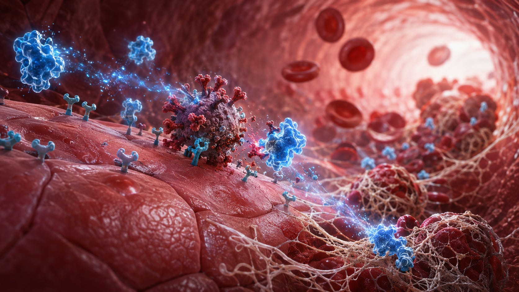
Public discourse about COVID-19 vaccine safety has largely stayed fixed on what regulators call “adverse events of special interest”: myocarditis, blood clots, anaphylaxis — the acute, countable, VAERS-reportable tip of the iceberg. But a growing peer-reviewed literature, built quietly across 2021–2026 by cardiologists, biochemists, and vascular researchers on several continents, describes something submerged and much larger: a spike protein that does not behave the way it was assumed to behave — that travels beyond the injection site, that can persist in blood, lymph tissue, and monocytes for weeks to months, and that appears capable of directly damaging blood vessels and distorting clot formation independent of any live virus at all.
What follows is the evidence: what spike protein does, where vaccine-derived material has been detected, how abnormal clotting enters the picture, and what the published literature shows about nattokinase.
Part I: The biology of the submerged mass
Spike protein is not an inert vaccine ingredient — it is a signaling molecule
The founding assumption behind every spike-based vaccine — whether delivered by mRNA, viral vector, or protein subunit — is that the spike protein functions primarily as an antigenic target. Independent laboratory research established that spike itself can also participate in biologically active signaling.
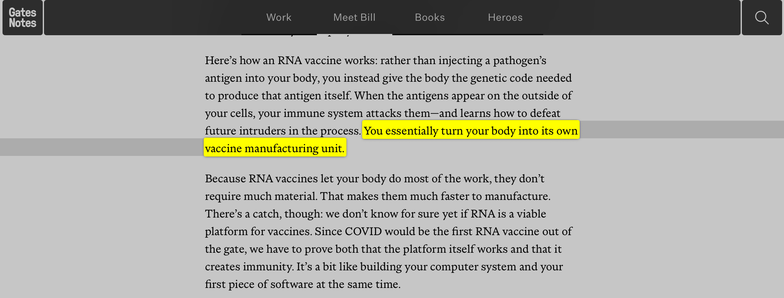
Bill Gates described the mRNA mechanism plainly in April 2020: rather than injecting a pathogen’s antigen, the platform gives the body genetic instructions to produce it, turning the body “into its own vaccine manufacturing unit.” Unlike a conventional vaccine that delivers a preformed antigen, the mRNA platform instructs the recipient’s own cells to manufacture the antigen. The relevant biological question, therefore, is what biologically active spike can do once produced.
Yuichiro Suzuki (Georgetown University Medical Center) and Sergiy Gychka (Bogomolets National Medical University) reviewed evidence in a peer-reviewed 2021 paper that spike protein binding to ACE2 triggers intracellular signaling cascades in human cells on its own, independent of viral replication — and explicitly raised the question of what this could mean for a vaccine strategy built around making the human body manufacture that same protein (Suzuki & Gychka, Vaccines, 2021).
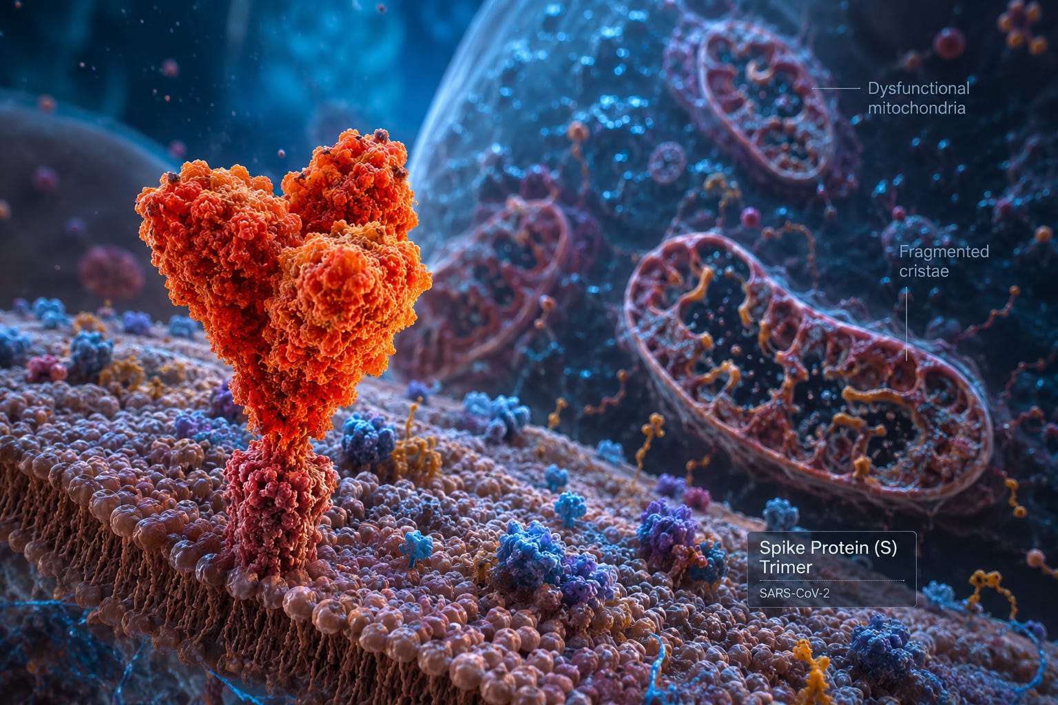
The most consequential single study in this literature came from a Salk Institute–UC San Diego–Xi’an Jiaotong University collaboration. Using a pseudovirus engineered to display spike protein but incapable of causing actual infection — meaning no other viral component was present — the team found that spike protein alone damages vascular endothelial cells by binding ACE2 and downregulating it, which in turn impairs mitochondrial function, increases reactive oxygen species, and shifts cell metabolism toward glycolysis (Lei et al., Circulation Research, 2021).
Lei et al. studied viral spike displayed on a nonreplicating pseudovirus, not vaccine-produced spike, so the experiment does not by itself establish vaccine injury. Salk’s own release emphasized that distinction. But the finding establishes something fundamental: spike itself can participate directly in vascular injury without viral replication.
A 2023 study extended the mechanism to heart muscle, finding that spike protein exposure directly impairs cardiomyocyte mitochondrial function — a proposed contributor to COVID-19-associated cardiac injury (Huynh et al., Cells, 2023). A 2023 review synthesized the broader literature under the term “spikeopathy,” arguing that viral and vaccine-induced spike expression can produce overlapping pathology (Parry et al., Biomedicines, 2023).
A central counterargument is that vaccine-encoded spike is structurally stabilized by proline substitutions and produced transiently at lower levels than spike generated during infection (FactCheck.org, 2021; Stanford Medicine, 2023). That addresses degree and duration. It does not erase the underlying biological point established by Lei and Suzuki: spike itself is biologically active rather than inert.
Where does it actually go? Biodistribution and persistence
The second pillar of the “submerged iceberg” thesis is that vaccine-derived material does not remain confined to the injection site and does not disappear as quickly as early public messaging implied.
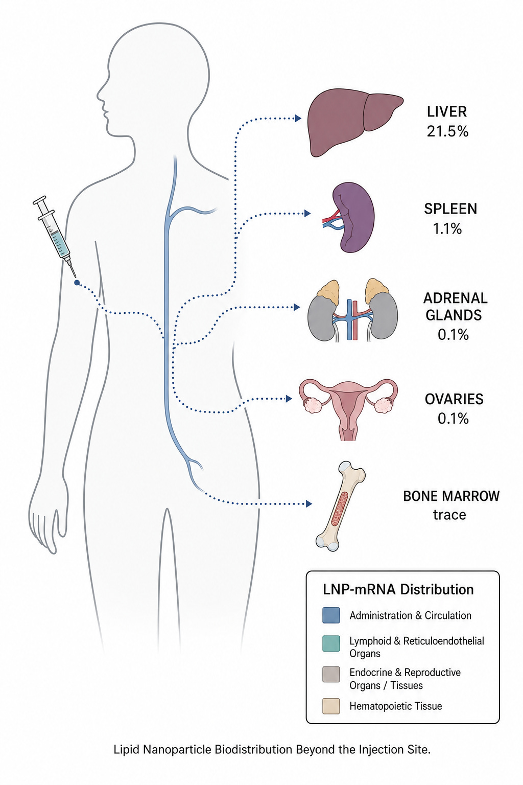
Pfizer’s nonclinical biodistribution data, submitted to Japan’s Pharmaceuticals and Medical Devices Agency (PMDA), is the primary regulatory source. In the radiolabeled-LNP rat study, the injection site held the greatest concentration at every time point; the liver was the dominant secondary depot, with measurable distribution to the spleen, adrenal glands, ovaries, and other tissues (PMDA Report, 2021; EMA assessment). A 2024 peer-reviewed review of RNA-vaccine biodistribution literature independently discusses the same systemic distribution pattern (Pateev et al., Biomedicines, 2024).
Using a single-molecule array (Simoa) assay roughly 1,000-fold more sensitive than standard antigen tests, Ogata et al. detected circulating spike-related protein in plasma as early as day one after vaccination, generally clearing within about two weeks as antibodies developed (Clinical Infectious Diseases, 2021).
A separate study found circulating exosomes — extracellular vesicles — carrying spike protein on their surface, first detectable on day 14, increasing after a booster dose, and declining in parallel with antibodies at around four months (Bansal et al., Journal of Immunology, 2021). The authors interpreted these spike-bearing exosomes as part of the vaccine’s immune mechanism, not as evidence of toxicity. The relevant observation remains: spike-bearing extracellular vesicles were measurable for months. The paper later generated a published methodological exchange (Somiya, J Immunol, 2022, and the authors’ response).
A large Stanford-led team separately found vaccine mRNA and spike antigen persisting within lymph-node germinal centers for up to eight weeks in some individuals (Röltgen, Boyd et al., Cell, 2022). A Danish study using ultrasensitive PCR detected vaccine mRNA sequences in blood plasma for up to 28 days post-vaccination in a subset of participants (Samaniego Castruita et al., APMIS, 2023).
These findings measure different compartments — free plasma protein, exosome-bound protein, circulating mRNA, and lymphoid-tissue-resident antigen — and therefore describe different clearance timelines. Taken together, they make the simplistic claim that vaccine-derived material uniformly “clears within days” untenable.
One mechanistic reason for prolonged expression is built into the platform itself: both authorized mRNA vaccines substitute N1-methylpseudouridine (m1Ψ) for uridine, a modification used specifically to reduce innate immune sensing and degradation, thereby improving translation and persistence (Nance & Meier, ACS Central Science, 2021). That is part of the published pharmacological rationale for the platform.
The clotting connection: how spike protein rewires fibrin
This is the mechanistic bridge that makes nattokinase relevant at all, so it deserves the most careful treatment.
A South African team led by Etheresia Pretorius (Stellenbosch University) and Douglas Kell (University of Liverpool) has built, since 2021, one of the more rigorous bodies of peer-reviewed work in this entire space. Their foundational experiment exposed platelet-poor plasma from healthy volunteers to recombinant spike protein S1 in vitro and found it induced anomalous, amyloid-containing fibrin(ogen) clots — structurally abnormal and markedly resistant to normal enzymatic breakdown compared to healthy clots (Grobbelaar, Venter, Pretorius et al., Bioscience Reports, 2021). Their follow-up work formalized this into the “fibrinaloid microclot” model of Long COVID: persistent, amyloid-structured clots that can physically obstruct capillaries, impair oxygen exchange, and entrap other plasma proteins (Kell, Laubscher & Pretorius, Biochemical Journal, 2022). A 2024 methodological paper reports these same fibrin amyloid microclots have separately been associated with disseminated intravascular coagulation and mortality in intensive-care COVID-19 patients elsewhere in the literature (Kell, Khan & Pretorius, Research and Practice in Thrombosis and Haemostasis, 2024).
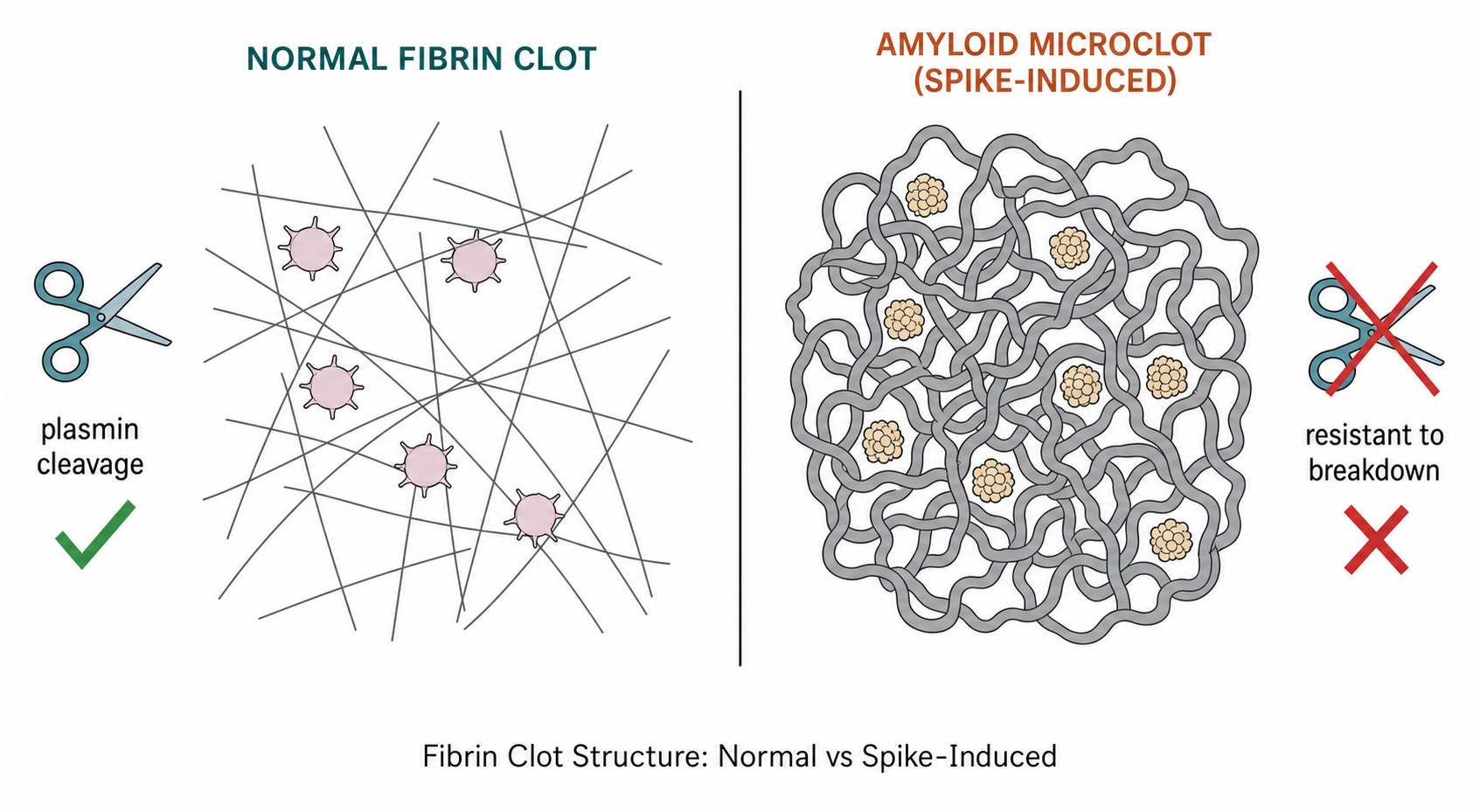
Most relevant here, a 2026 proteomic study from an overlapping Pretorius research group reported amyloid deposits and coagulopathy-associated inflammatory signatures in a combined post-vaccination/post-infection syndrome cohort (Waters, Vlok, Pretorius et al., Frontiers in Cellular and Infection Microbiology, 2026). Because the cohort combines both groups, it cannot establish that the findings are uniquely vaccine-induced, and independent replication is still needed.
There is also a specific mechanistic account of how spike protein activates platelets directly: through the TMEM16F ion channel, driving procoagulant activity independent of the classical clotting cascade (Cappelletto, Giacca et al., Frontiers in Cardiovascular Medicine, 2023). The same paper’s post-mortem cohort found lung microthrombosis in 83% of ICU COVID-19 patients — important context, though describing infection rather than vaccination.
VITT should not be confused with the mechanism discussed here. It is a distinct PF4-antibody-mediated syndrome associated predominantly with adenoviral-vector vaccines rather than mRNA vaccination (Cines & Greinacher, Blood, 2023).
The cardiac signal
Myocarditis following mRNA vaccination is now one of the clearest quantified vaccine safety signals. Its distribution is highly nonuniform: risk is concentrated in younger males, particularly after second doses, and is higher after Moderna than Pfizer in several large datasets.
CDC’s Advisory Committee on Immunization Practices, reviewing VAERS data through June 2021, found the signal concentrated by age and sex: roughly 40.6 myocarditis cases per million second doses in males aged 12–29, versus 4.2 per million in females of the same age, and roughly 1–2 per million in adults over 30 (WHO GACVS, 2021; CDC MMWR, 2021). Israel’s national health data corroborated the pattern (Mevorach et al. and Witberg et al., both NEJM, 2021).
The largest and most granular dataset comes from a pooled Nordic cohort of 23.1 million residents (Karlstad et al., JAMA Cardiology, 2022). Among males aged 16–24 receiving two doses of the same vaccine, the adjusted incidence rate ratio after a second dose was 5.31 for Pfizer-BioNTech and 13.83 for Moderna. In absolute terms, that corresponds to 5.55 excess myocarditis events per 100,000 vaccinees after a second Pfizer dose and 18.39 per 100,000 after a second Moderna dose.
A re-analysis of the original Pfizer and Moderna phase III trial data using the WHO Brighton Collaboration adverse-event framework also found a statistically significant excess of serious adverse events of special interest in Pfizer’s vaccine arm relative to placebo (Fraiman et al., Vaccine, 2022).
SARS-CoV-2 infection itself also increases myocarditis risk and in some population-level analyses produces a larger aggregate signal than vaccination (Patone et al., Circulation, 2022). That does not erase the vaccine signal; it defines it more precisely. Vaccine-associated myocarditis is a real, demographically concentrated adverse event, not a uniform risk across the population.
Part II: Enter nattokinase
What it actually is
Nattokinase (NK) is a subtilisin-family serine protease discovered in 1980 by Japanese researcher Hiroyuki Sumi, then at the University of Chicago Medical School, and formally described in the literature in 1987 (Sumi et al., Experientia, 1987). It comes from natto — the sticky fermented-soybean dish that has been a staple of the Japanese diet for centuries — and is produced by fermenting soybeans with Bacillus subtilis var. natto. The resulting 275-amino-acid enzyme is closely homologous to subtilisin E, placing it squarely in a well-characterized family of bacterial proteases with a classic serine-protease catalytic triad (Weng et al., International Journal of Molecular Sciences, 2017).
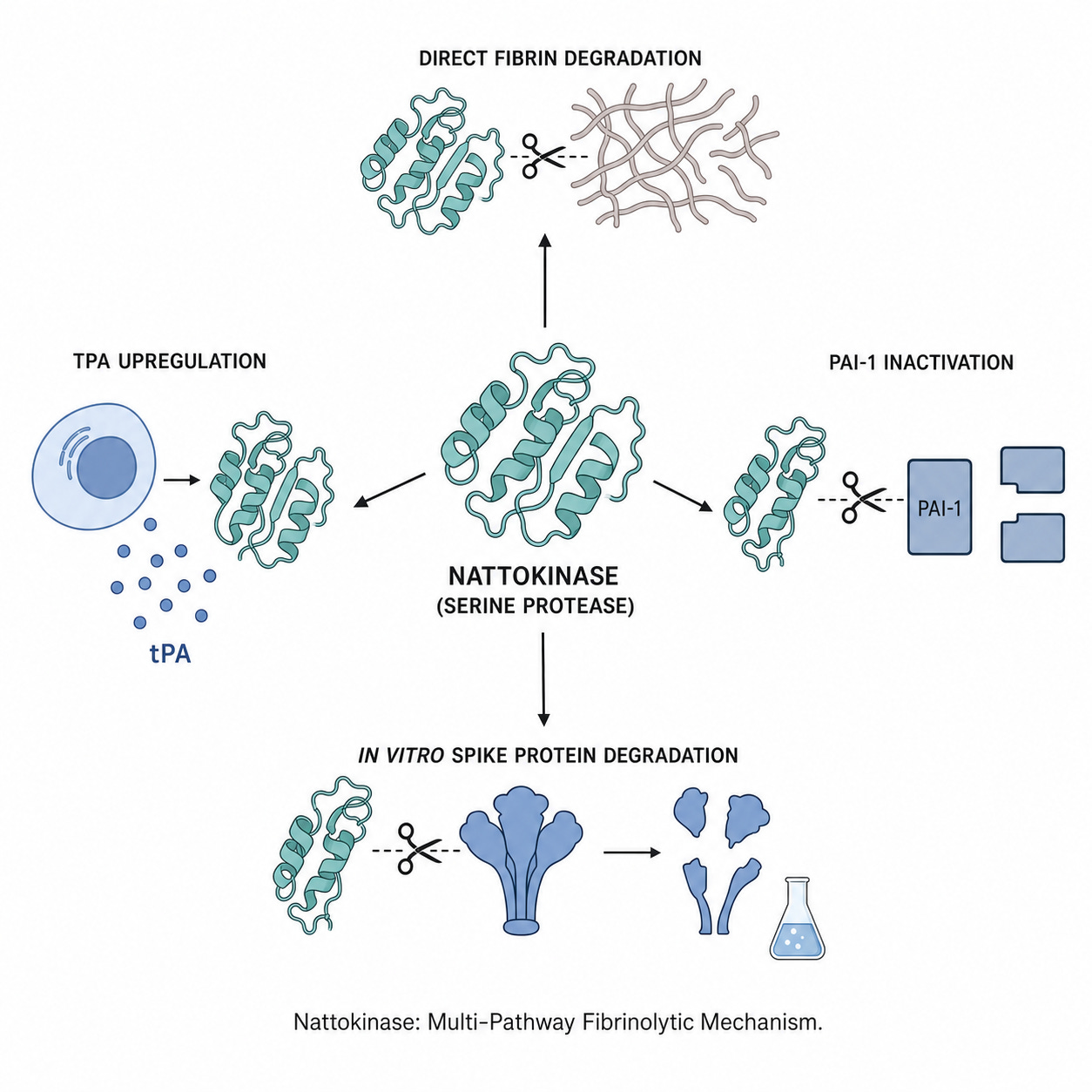
NK’s cardiovascular relevance comes from a multi-pronged mechanism, not a single trick. It cleaves cross-linked fibrin directly, in a manner analogous to the body’s own plasmin (Sumi et al., 1987). It promotes the release of tissue plasminogen activator (tPA) and converts inactive prourokinase into active urokinase, amplifying the body’s own clot-dissolving cascade rather than simply substituting for it (Weng et al., 2017). And in perhaps its most elegant documented mechanism, it directly cleaves and inactivates PAI-1 — plasminogen activator inhibitor type 1, the molecular brake that normally restrains fibrinolysis (Urano et al., Journal of Biological Chemistry, 2001). In the same study, tPA-induced clot lysis was substantially greater in the presence of NK when PAI-1 was present than when it was absent — direct biochemical evidence that disabling the brake, not just cutting the rope, is a meaningful part of how this enzyme works.
None of this is speculative or “alternative” biochemistry. It is published, peer-reviewed enzymology that any hematologist would recognize, even if most have never been asked to consider a food-derived enzyme as a clinically relevant fibrinolytic agent.
Does it actually degrade spike protein?
This is the load-bearing question, and it deserves more precision than it receives in either direction — from those who treat it as settled and from those who dismiss it as fabrication.
Two independent, peer-reviewed, in vitro studies exist. Both hold up under scrutiny, with important caveats attached to each.
The first, more frequently cited, is a 2022 study in Molecules. Researchers transfected human embryonic kidney (HEK293) cells with plasmids encoding full-length SARS-CoV-2 spike protein, an RBD-GFP fusion construct, or human ACE2, then exposed cell lysates and intact live cells to nattokinase. Results were dose- and time-dependent: nattokinase degraded spike protein at concentrations as low as 7.8 ng/mL in a four-fold dilution series, required at least 60 minutes of exposure to show an effect at 1 µg/mL, and also caused loss of the RBD and, separately, the human ACE2 receptor protein itself at 7.5 µg/mL. Heating the enzyme to 100°C or adding serine-protease inhibitors abolished the effect entirely, which is the correct control confirming a genuine enzymatic mechanism rather than an assay artifact (Tanikawa et al., Molecules, 2022).
One limitation matters: the same study found nattokinase also degraded GAPDH under identical conditions. Nattokinase is not a spike-specific enzyme; it is a broad protease capable of degrading spike when the two encounter one another at sufficient concentration and exposure time.
Three co-authors were affiliated with Contek Life Science Co., Ltd., the nattokinase manufacturer whose product was tested, despite the paper declaring no conflict of interest.
The second study is more reassuring on the conflict question, coming from an unaffiliated Japanese academic consortium (Tokyo University of Agriculture and Technology and the National Institute of Technology) with no apparent commercial nattokinase ties. Using actual live SARS-CoV-2 isolated from a Diamond Princess patient — not recombinant protein fragments — the team found that whole natto extract fully inhibited viral infection of cultured cells, and on Western blot showed the extract proteolytically degraded the receptor-binding domain, including a variant construct carrying the N501Y mutation found in the Alpha variant. Again, heat treatment and protease inhibitors reversed the effect (Oba et al., Biochemical and Biophysical Research Communications, 2021). The caveat here is different: this study used whole natto extract, not purified isolated nattokinase, so it cannot be said with certainty that nattokinase specifically — as opposed to some other bioactive component of fermented natto — was responsible, though nattokinase is natto’s dominant and best-characterized protease.
Because these two studies come from different groups, use different methods, and were published a year apart, their convergence is meaningful. This is real, reproducible, peer-reviewed biochemistry.
The crucial question: does orally consumed nattokinase reach systemic circulation intact?
The spike-degradation experiments applied nattokinase directly to cells or proteins. A swallowed capsule is a different biological problem.
Nattokinase is a roughly 275-amino-acid protein, and proteins of that size are generally expected to undergo substantial digestion in the gastrointestinal tract. Yet there is evidence of systemic biological activity after oral dosing. Rodent studies have detected intact enzyme crossing the intestinal tract, and in humans, a single oral 2,000 FU dose produced measurable changes in D-dimer and fibrin/fibrinogen degradation products within hours — downstream markers of systemic fibrinolysis.
What has not yet been established is the circulating concentration of intact, catalytically active nattokinase in humans — or whether it reaches levels sufficient to reproduce the spike-degradation effects observed in vitro. That is the critical experiment still missing. But it does make sense that manufacturers of dietary supplements use an enterically coated capsuel in order to ensurce survival through the stomach for higher absorption through the small intestine.
The cardiovascular record — and the dose question
Long before COVID-19, nattokinase had accumulated a genuine if uneven clinical trial record for cardiovascular disease.
The foundational human RCT — eight weeks, placebo-controlled, 86 pre-hypertensive and stage-1-hypertensive adults — found that 2,000 FU/day lowered systolic blood pressure by roughly 5.55 mmHg versus placebo (Kim et al., Hypertension Research, 2008).
The most methodologically rigorous nattokinase trial conducted to date — three years, 265 patients, double-blind, randomized, placebo-controlled, using carotid intima-media thickness as its endpoint — found no effect on atherosclerosis progression at 2,000 FU/day in healthy, low-risk adults (Hodis et al., Clinical Hemorheology and Microcirculation, 2021). It is a strong null result and it matters.
A retrospective study of 1,062 participants reported that at 10,800 FU/day over twelve months, carotid intima-media thickness and plaque area fell substantially (up to a 36% reduction) alongside lipid improvements, while an internal comparison group at 3,600 FU/day showed no significant benefit (Chen et al., Frontiers in Cardiovascular Medicine, 2022).

One co-author was affiliated with Sungen Bioscience Co. Ltd., and the paper carries a published corrigendum. A separate 76-patient randomized comparison against simvastatin at 6,500 FU/day reported greater carotid plaque reduction with nattokinase despite less aggressive cholesterol lowering (Ren et al., Zhonghua Yi Xue Za Zhi, 2017). I wrote about this landmark research last year, and it inspired me to create my own version, given it was difficult to find products with a 10,800 dosage on the market, and certainly not one whose quality I could personally ensure.
Hodis and Chen point in opposite directions, but dose is not the only difference. Hodis was randomized, double-blind and placebo-controlled in low-risk adults; Chen was retrospective and non-randomized in hyperlipidemic patients. The strongest evidence for a dose effect therefore comes not from comparing the two studies, but from Chen’s own internal comparison: 3,600 FU/day showed no significant plaque benefit while 10,800 FU/day did.
That makes a dose-response effect plausible, but not yet established by randomized evidence. A pooled 2024 meta-analysis reports significant blood-pressure benefit across doses but no significant lipid benefit at low doses over short durations, which is at least consistent with a dose-dependent picture.
Our product provides 10,800 FU per serving, matching the dose used in Chen and substantially exceeding the 2,000-FU dose tested in Hodis. We selected that dose based on the existing literature, while recognizing that a definitive randomized dose-ranging trial has not yet been performed.
Part III: The protocols people are already using
The Front Line COVID-19 Critical Care Alliance lists nattokinase (100–200 mg, twice daily) as a first-line agent in its I-RECOVER post-vaccine-syndrome protocol, describing it as a fibrinolytic and antiplatelet agent targeting abnormal clotting and citing the Tanikawa 2022 study among its mechanistic foundations (FLCCC I-RECOVER, 2023). Their protocol is well worth reading.
The most direct academic articulation of the “spike detox” rationale comes from a 2023 paper in the Journal of American Physicians and Surgeons by Anastasia Kyriakopoulos, Greg Nigh, Peter McCullough, and MIT’s Stephanie Seneff. The authors propose that persistent spike protein may resist normal proteolytic disposal and drive chronic inflammation via NF-κB activation, and they catalogue nattokinase, ASPNJ, and serrapeptidase as candidate remedies. The paper discloses that McCullough is chief scientific officer of The Wellness Company, which markets a nattokinase-containing supplement.
Bromelain has its own smaller, mechanistically distinct evidence base. I’ve been indexing research on this remarkable pineapple-derived substance for almost two decades. You can view it on GreenMedInfo.com here. Serrapeptidase, although commonly bundled into “systemic enzyme” protocols, has essentially no known direct spike-degradation study behind it.
What remains unproven
No controlled human trial has yet shown that nattokinase clears vaccine-derived spike from human tissue or improves post-vaccination syndrome. The clinical rationale therefore rests on the convergence of established fibrinolytic activity, experimental spike degradation, and clinical experience — not completed efficacy trials.
That distinction matters. The enzymology showing that nattokinase can cut spike protein in vitro is real and peer-reviewed. Whether oral nattokinase reproduces that effect inside the human body remains the central clinical question.
Part IV: Safety — this is a drug-grade enzyme, not a vitamin
Nattokinase’s fibrinolytic activity is precisely why it is biologically interesting and why it demands respect. Foundational toxicology found no adverse effects in a 28-day human trial at 10 mg/kg/day, and animal studies found no mutagenic or clastogenic effects even at doses roughly 100-fold higher than typical human intake (Lampe & English, Food and Chemical Toxicology, 2016).
Rare serious events have nevertheless been reported, particularly in people using anticoagulants or with substantial vascular risk: a cerebellar hemorrhage in a stroke patient who added nattokinase to daily aspirin (Chang et al., Internal Medicine, 2008); mechanical heart-valve thrombosis in a patient who substituted nattokinase for prescribed warfarin (Elahi et al., Proceedings (Baylor University Medical Center), 2015); and a fatal case of hemoperitoneum in an elderly woman taking over-the-counter nattokinase (Ramachandran et al., Cureus, 2021).
Counterbalancing these, a real-world study of 153 vascular-surgery patients — some concurrently taking prescription anticoagulants — reported symptom improvement with no recorded adverse drug reactions, while emphasizing the need for monitoring when nattokinase is combined with blood-thinning therapy (Gallelli et al., Nutrients, 2021).
Because nattokinase promotes fibrinolysis, concurrent use with anticoagulant or antiplatelet drugs can increase bleeding risk. People taking warfarin, DOACs, aspirin or other antiplatelet agents; those with bleeding disorders, prior hemorrhagic stroke or cerebral microbleeds; people with mechanical heart valves; and those approaching surgery should use it only under appropriate clinical supervision. This is why it is always advisable to consult a licensed health care practitioner or medical herbalist before using dietary supplements while already on medications, of any kind.
Most reassuring safety data come from products dosed at or near 2,000 FU. High-dose studies report no serious adverse events, but they are fewer, smaller, and mostly not randomized. If higher doses produce stronger biological effects, greater bleeding risk must also be considered, not excluding over-the-counter drugs like aspirin, whose side effects I have documented extensively. That is especially relevant to high-dose preparations, including ours at 10,800 FU per serving.
Why hasn’t the definitive trial been run?
If a fermented-soybean or garbanzo bean enzyme can degrade spike protein in vitro and is already being used by large numbers of people for post-COVID and post-vaccination symptoms, why is there still no large, funded, placebo-controlled trial testing it?
The funding incentives are badly misaligned. Nattokinase is not a patentable new molecular entity attractive to pharmaceutical development, while supplement manufacturers can already market nattokinase without financing the kind of multimillion-dollar outcomes trial that could produce either a spectacular validation or an expensive null result.
The supplement industry does fund research — Contek Life Science co-authored one nattokinase study and Sungen Bioscience another — but the literature remains dominated by mechanistic work, smaller studies, retrospective analyses, and a rigorous null RCT at a lower dose.
The predictable result is the evidence base we have today: compelling mechanistic findings, several small or observational clinical studies, one strong null randomized trial at 2,000 FU/day, and no definitive outcomes trial addressing post-vaccination or post-COVID syndromes.
The absence of that trial does not negate the biological findings already on the table. It identifies the experiment that still needs to be done.
The bottom line
The biological foundation is no longer trivial. Spike protein is biologically active. It can injure vascular endothelium, disrupt mitochondrial function, and alter fibrin structure. Vaccine-derived mRNA and antigen have been detected outside the injection site and for substantially longer periods than the simplest early public messaging suggested.
Nattokinase enters this picture for two independent reasons. It is a well-characterized fibrinolytic enzyme with decades of cardiovascular research behind it, and independent laboratory studies have demonstrated that proteolytic preparations containing nattokinase can degrade SARS-CoV-2 spike protein or its receptor-binding domain under experimental conditions.
The missing link is human clinical confirmation. We still need to know how much catalytically active nattokinase reaches systemic circulation after oral dosing, whether it materially reduces persistent spike or abnormal fibrin in vivo, which patients benefit, and at what dose.
Those are not reasons to dismiss the existing evidence. They are the experiments that should have been performed years ago.
Millions of people have already been left to navigate post-COVID and post-vaccination symptoms without a coherent research program addressing this specific hypothesis. When a centuries-old fermented food produces an enzyme with documented fibrinolytic activity and experimental capacity to degrade the very protein at the center of this pathology, the appropriate scientific response is neither credulity nor ridicule.
It is to test it.
This article is for educational purposes and is not medical advice. Nattokinase is a potent fibrinolytic and antiplatelet agent. Do not start, stop, or combine it with any medication — especially anticoagulants, antiplatelet drugs, or in preparation for surgery — without consulting a qualified healthcare provider who knows your full medical history. The author has a financial interest in a company that sells a nattokinase-containing supplement, disclosed in full at the top of this article.
Key citations
1. Suzuki YJ, Gychka SG. “SARS-CoV-2 Spike Protein Elicits Cell Signaling in Human Host Cells: Implications for Possible Consequences of COVID-19 Vaccines.” Vaccines. 2021;9(1):36. PMC7827936
2. Lei Y, Zhang J, Schiavon CR, et al. “SARS-CoV-2 Spike Protein Impairs Endothelial Function via Downregulation of ACE2.” Circulation Research. 2021;128(9):1323–1326. DOI
3. Huynh TV, et al. “Spike Protein Impairs Mitochondrial Function in Human Cardiomyocytes.” Cells. 2023;12(6):877. PMC10046940
4. PMDA. “Report on the Deliberation Results” (Comirnaty biodistribution data). 2021. PDF
5. Ogata AF, Cheng CA, et al. “Circulating SARS-CoV-2 Vaccine Antigen Detected in the Plasma of mRNA-1273 Vaccine Recipients.” Clinical Infectious Diseases. 2021. PMC8241425
6. Bansal S, Perincheri S, et al. “Circulating Exosomes with COVID Spike Protein Are Induced by BNT162b2 Vaccination.” Journal of Immunology. 2021;207(10):2405–2410. PMID 34654691
7. Röltgen K, Boyd SD, et al. “Immune imprinting, breadth of variant recognition, and germinal center response in human SARS-CoV-2 infection and vaccination.” Cell. 2022. PMID 35148837
8. Nance KD, Meier JL. “The Role of N1-Methylpseudouridine in COVID-19 Vaccines.” ACS Central Science. 2021. PMC8043204
9. Grobbelaar LM, Venter C, Pretorius E, et al. “SARS-CoV-2 spike protein S1 induces fibrin(ogen) resistant to fibrinolysis.” Bioscience Reports. 2021;41(8). PMC8380922
10. Kell DB, Laubscher GJ, Pretorius E. “A central role for amyloid fibrin microclots in long COVID/PASC.” Biochemical Journal. 2022;479(4):537–559. PMC8883497
11. Cappelletto A, Giacca M, et al. “SARS-CoV-2 Spike Protein Activates TMEM16F-Mediated Platelet Procoagulant Activity.” Frontiers in Cardiovascular Medicine. 2023. Article
12. Cines DB, Greinacher A. “Vaccine-induced immune thrombotic thrombocytopenia.” Blood. 2023. PMC9870607
13. Karlstad Ø, Hviid A, Ljung R, et al. “SARS-CoV-2 Vaccination and Myocarditis in a Nordic Cohort Study of 23 Million Residents.” JAMA Cardiology. 2022. Article
14. Patone M, et al. “Risk of myocarditis after sequential doses of COVID-19 vaccine and SARS-CoV-2 infection.” Nature Medicine. 2022. Article
15. Fraiman J, Erviti J, Jones M, Greenland S, Doshi P, et al. “Serious Adverse Events of Special Interest Following mRNA COVID-19 Vaccination in Randomized Trials in Adults.” Vaccine. 2022. PMID 36055877
16. Sumi H, Hamada H, Tsushima H, et al. “A novel fibrinolytic enzyme (nattokinase) in the vegetable cheese Natto.” Experientia. 1987;43:1110–1111. PMID 3478223
17. Urano T, Ihara H, Umemura K, et al. “The profibrinolytic enzyme subtilisin NAT cleaves and inactivates plasminogen activator inhibitor type 1.” Journal of Biological Chemistry. 2001;276:24690–24696. PMID 11325965
18. Tanikawa T, Kiba Y, Yu J, et al. “Degradative Effect of Nattokinase on Spike Protein of SARS-CoV-2.” Molecules. 2022;27(17):5405. PMC9458005
19. Oba M, Rongduo W, Saito A, et al. “Natto extract, a Japanese fermented soybean food, directly inhibits viral infections including SARS-CoV-2 in vitro.” Biochemical and Biophysical Research Communications. 2021;570:21–25. Article
20. Kim JH, Gum SN, Paik JK, et al. “Effects of Nattokinase on Blood Pressure: A Randomized, Controlled Trial.” Hypertension Research. 2008;31(8):1583–1588. Article
21. Hodis HN, Mack WJ, Meiselman HJ, et al. “Nattokinase atherothrombotic prevention study.” Clinical Hemorheology and Microcirculation. 2021;78(4). DOI
22. Chen H, Chen J, Zhang F, et al. “Effective management of atherosclerosis progress and hyperlipidemia with nattokinase: A clinical study with 1,062 participants.” Frontiers in Cardiovascular Medicine. 2022;9:964977. PMC9441630
23. Ren N, Chen H, Li Y, McGowan E, Lin Y. “A clinical study on the effect of nattokinase on carotid artery atherosclerosis and hyperlipidaemia.” National Medical Journal of China. 2017;97:2038–2042. Summary, PMC6043915
24. Lampe BJ, English JC. “Toxicological assessment of nattokinase derived from Bacillus subtilis var. natto.” Food and Chemical Toxicology. 2016;88:87–99. Cited via PMC6043915
25. Kyriakopoulos AM, Nigh G, McCullough PA, Seneff S. “Proteolytic Targets for SARS-CoV-2 Spike Protein Degradation: Hope for Systemic Detoxification.” Journal of American Physicians and Surgeons. 2023;28(3):86–89. PDF
26. McCullough PA, Wynn J. “Clinical Rationale for SARS-CoV-2 Base Spike Protein Detoxification in Post COVID-19 and Vaccine Injury Syndromes.” Journal of American Physicians and Surgeons. 2023;28(3):91–93. PDF
27. Front Line COVID-19 Critical Care Alliance. “I-RECOVER: Management of Post-Vaccine Syndrome.” July 24, 2023. PDF
28. Chang YY, Liu JS, Lai SL, Wu HS, Lan MY. “Cerebellar hemorrhage provoked by combined use of nattokinase and aspirin.” Internal Medicine. 2008;47(5):467–469. PMID 18310985
29. Elahi MM, Choi CH, Konda S, Shake JG. “Consequence of patient substitution of nattokinase for warfarin after aortic valve replacement.” Proceedings (Baylor University Medical Center). 2015;28(1):81–82. PMC4264722
30. Ramachandran L, Aqeel A, Jafri A, Sidhu Y, Mohamed Djirdeh T. “Nattokinase-Associated Hemoperitoneum in an Elderly Woman.” Cureus. 2021;13(12):e20074. PMID 35003944
31. Gallelli G, et al. “Data Recorded in Real Life Support the Safety of Nattokinase in Patients with Vascular Diseases.” Nutrients. 2021;13(6):2031. PMC8231931
32. FactCheck.org. “Posts Push Unproven ‘Spike Protein Detoxification’ Regimen.” 2023. Article
Will Sprouts have coupons for your Nattokinase soon? I asked Sprouts if they had any because sometimes they do with new products. They said no and contact the company. I guess that is you! 😃
While there are no RCT's, people have been claiming to use nattokinaise for years to reduce spike protein. As there are now tests for spike proteins, isn't there anecdotal info from people who have measured, taken natto, then measured again?